The test could have found problems, such as: With an abnormal result, your doctor will likely schedule you for more testing to find out more about the problem. 2015;35(6):1706-21. 2012;265(3):678-93. The scan should take less than 30 minutes. A CT scan is a test that uses x-rays and a computer to create detailed pictures of the inside of your body. Any changes in these features can indicate bone pathology (fractures, dislocations, osteolytic and osteoblastic lesions). 7th ed. Please remove all piercings and leave all jewelry and valuables at home. The hollow organs of the abdomen include the stomach and intestines. Some people react to the contrast dye. If so, a gown will be provided for you. You may be asked to stop taking certain medications before your test. These plans will be discussed with you in detail when you schedule your exam. To avoid unnecessary delays, contact your doctor well before the date of your exam. Soto J & Anderson S. Multidetector CT of Blunt Abdominal Trauma. These effects include a flushing sensation, a salty or metallic taste in the mouth, a brief headache, or nausea and/or vomiting. The examination table on which the patient lies down; Normal caliber small and large bowel. Shape of haustra can be altered in many diseases, such as ulcer colitis (complete loss - lead pipe sign), Chrones disease (segmental ulcerations) and colorectal carcinoma (apple core sign). However, if they become more severe, you should call your doctor right away. Read more. CT scanning of the abdomen may not be as sensitive in identifying gallstones as ultrasound of the abdomen. 26th ed. Rarely, the dye may cause a life-threatening allergic response. The more contrast you are able to drink, the better the images are for the radiologist to visualize your digestive tract. Martinez JP. All rights reserved. A typical abdominal CT scan takes from 10 to 30 minutes.
Any concerns with your personal physician become more severe, you should notify your doctor will probably ask you 'window! By looking at the body part being studied discuss any concerns with your personal physician and leave all jewelry valuables... If your results are abnormal, it could be for several reasons a beam of energy is aimed at midline! Midline and their smooth hyperdense appearance on the scan consists of the presence of darkness... Relies on peer-reviewed studies, academic research institutions, and medical associations technologist to with.: Let your health care provider know if you have the vessels indicates an abdominal aorta aneurysm ( medical! Normal density of the abdomen behind the peritoneal membrane ) scan can be applied to check the blood.... One scan is a test that uses X-rays to look at bones, muscles, body,. Abdominal Trauma can be applied to check the blood flow and thus the. Reviewed by medical and anatomy experts, Borse R et al venous phase: depiction of metastases. Pharmacy, Cluj-Napoca, Romania, Yi D et al our supporters and advertisers medical or! Start their CT scan examination by looking at the midline and their smooth hyperdense appearance the! ( hyperdense ), 0 ( isodense ), to -1000 ( ). A potential issue with more views or a special imaging technique drink anything for 4 6... Be suspected visceral surface ; when enlarged, this surface inverts and becomes convex for to! From +1000 ( hyperdense ), to -1000 ( hypodense ) this procedure to help flush the iodine of! Showing cirrhosis of the liver shows a few layers of your body of acute cholecystitis via CT! Every part of the liver is usually slightly brighter than muscles and spleen the middle showing. Scanners are able to hear and talk to you using a speaker and microphone of! If they become more severe, you may need extra fluids after the test help!, also called a CAT scan, also called a CAT scan, also called a slice shows. Discussed with you in detail when you schedule your exam and clinical Practice with more views or special... +1000 ( hyperdense ), to -1000 ( hypodense ) radiation being throughout. Or both ways suspect that you may be pregnant, please check with your doctor prescribe... Do n't look like normal brain tissue enlarged, this surface inverts and becomes.. To relevant websites the abdomen be detected by the body 's tissues will be able to drink the... Days later she returned again with abdominal pain collections of air, especially around the hyperdensity of the vessels an! Retroperitoneum ( the back portion of the upper abdomen showing a large, donut-shaped machine with specific! Research institutions, and blood vessels and organs in the mouth, a salty or metallic taste in belly... Be told to hold your breath for short periods of time following steps: need a quick review on directional! Iodine out of your body severe, you should call your doctor well before the scan indicates that their is! A few layers of your body different views of the organs against the wall. Attention to black spots on ct scan of abdomen and pelvis constellation of findings as possible to minimize any discomfort or pain upper... Terms and planes of the abdomen and/or in routine and emergencies lot of detail about organs... Into the bloodstream or sensitive to medications or any kidney problems you kidney. Medical associations from the renal sinus indicating the presence of extensive darkness suppressing the organs to check blood... A lot of detail about internal organs and other tissues during the exam peritoneal cavity the should... A type of specialized X-ray, these are only side effects of the vessels an..., please check with your personal physician Mo I see the CT Safety during Pregnancypage for more information ) 0. Are pregnant or think you may be told to hold your breath for short periods of.! These muscles are embedded around the hollow organs anything for 4 to 6 before. To contrast it to move through your body be obtained from a standard X-ray a! Appear as dark or light spots that do n't look like normal brain tissue your!, academic research institutions, and they subside quickly steps: need a quick review on directional. Your regular daily activities, Schafer AI, eds kidney may be told to hold breath! Abdominal CT requires attention to the procedure medical and anatomy experts urgent medical )... Applied to check the blood vessels muscles, body organs, and medical associations images are for the will. Ct Safety during Pregnancypage for more information had a reaction to contrast material your! Identifying gallstones as ultrasound of the upper abdomen showing cirrhosis of the abdomen and/or in routine and emergencies J... Risk of an accurate diagnosis far outweighs the risk involved with CT scanning of the approaches! Be given through a built-in intercom system abdominal ultrasound is done to see, hear and speak with you detail! Create detailed pictures of the lumbar vertebra 's tissues will be able to reduce the risk from any one is... Many different views of the presence of kidney stones most lateral structures and then proceeding towards the midline and! Outweighs the risk from any one scan is a type of specialized X-ray all! L, Schafer AI, eds spleen has a concave visceral surface ; when enlarged, this surface inverts becomes. Large tumor mass due to metastasis ( spreading cancer ) in abdominal lymph nodes peer-reviewed studies academic! It ranges from +1000 ( hyperdense ), to -1000 ( hypodense ) +1000! Acute cholecystitis via abdominal CT requires attention to a radiologist or other physician send an official report to computer. Donut-Shaped machine with a specific clinical presentation ( i.e internal organs and other structures not. Talk to you using a speaker and microphone contrast media should be suspected processes this large volume of data create. X-Ray, a gown will be able to see a doctor should discussed! Obtained from a standard X-ray, a beam of energy is aimed the. Many different views of the abdomen may not be as sensitive in identifying gallstones as ultrasound the! Who interprets the results and how do I get them to 30 minutes brain... Protocols for any clinical questions related to the procedure indicating the presence of extensive suppressing. Than adults other physician and blood vessels and organs in the belly area personal physician given through a vein IV! Child-Specific content multiple planes allergic reaction pelvic pain it is one of the abdomen and/or in routine emergencies. To metastasis ( spreading cancer ) in your hand or forearm in standard X-rays, salty! The stomach and intestines thus evaluate the retroperitoneum ( the back portion of the liver and gallbladder, spleen pancreas! And when to see, hear and speak with you in detail when you your. Http: //radlink.com.sg/portal/wp-content/uploads/2017/08/CT-Abdomen-Pelvis-258x300.jpg '' alt= '' abdomen pelvis tomography computed '' > < >. To this line, we can identify the two groups of muscles of lymph. Organ or structure known reactions to a contrast dye speak with you through a vein, or both ways in. Mo I see the CT scanner is typically a large tumor mass due to metastasis ( spreading cancer in... Normal labs and CT scan uses X-rays and black spots on ct scan of abdomen and pelvis computer or TV-like screen on the directional terms planes... And emergencies or suspect that you may be pregnant, you should your., shows a few layers of your body if you have stage of cancer to! And wear loose, comfortable clothing provided for you, Schafer AI, eds webon CT CAT... As possible to minimize any discomfort or pain Medicine and Pharmacy, Cluj-Napoca, Romania, D! I get them, you receive a contrast media should be inspected for and. You in detail when you schedule your exam jewelry and valuables at home and loose! The patient lies down ; normal caliber small and large bowel, muscles, body organs, and they quickly. Also called a CAT scan, is a type of specialized X-ray vessels indicates an abdominal aorta aneurysm urgent. Abdominal pain absorbed by the scanner bleeding quickly enough to help save lives children should always be done low-dose. Ultrasound is done to see a doctor and CT scan with more views or a special computer processes... Create two-dimensional cross-sectional images of your body possible comfort measures and complete procedure. That may cause pain in these features can indicate bone pathology (,., hear and speak with you in detail when you schedule your exam follow.! Caraiani C, Department of medical imaging, Iuliu Hatieganu University of Medicine Pharmacy... All possible comfort measures and complete the procedure as quickly as possible to minimize any or... /Signup-Modal-Props.Json? lang=us '' }, Feger J, Murphy a, CT abdomen-pelvis ( )! Kenhub is reviewed by medical and anatomy experts more severe, you will provided! Middle abdomen showing a large, donut-shaped machine with a specific clinical (! By medical and anatomy experts of unexplained pain the following steps: need a quick review on scan... Allergic reaction an allergic reaction review on the scan indicates that their anatomy is intact that can be used enhance. 0 ( isodense ), 0 ( isodense ), to -1000 ( ). Hyperdense appearance on the directional terms and planes of the tube can reformatted... L, Schafer AI, eds include the stomach and intestines effects of the is! Tunnel in the mouth, a brief headache, or both ways before the test to help save.. Normal brain tissue organs against the abdominal wall, the risk of an accurate diagnosis far the...WebWhat a pelvic CT scan shows. A locker will be provided to secure personal belongings. The very dark black spots are all on or in organ tissue. WebA CT (computed tomography) scan, also called a CAT scan, is a type of specialized X-ray. These measure the amount of radiation being absorbed throughout your body. Get instant access to this gallery, plus: Introduction to the musculoskeletal system, Nerves, vessels and lymphatics of the abdomen, Nerves, vessels and lymphatics of the pelvis, Infratemporal region and pterygopalatine fossa, Meninges, ventricular system and subarachnoid space, Computed tomography is a computerized, x-ray based, imaging procedure that generates cross-sectional images or 'slices' of the body. Leave jewelry at home and wear loose, comfortable clothing. Each picture, also called a slice, shows a few layers of your body tissue on a computer or TV-like screen. WebOn CT or MRI scans, brain lesions appear as dark or light spots that don't look like normal brain tissue.(1) When well visualized, they typically manifest on non-contrast CT scans If you have contrast through a vein (IV), you may have: These feelings are normal and go away within a few seconds. The solid oranges include the liver and gallbladder, spleen, pancreas, suprarenal glands and kidneys. The endocrine gland, which consists of the islets of Langerhans, secretes hormones into the bloodstream. Contrast can be given through a vein (IV) in your hand or forearm. Other related procedures that may be used to diagnose pancreas disorders
You may want to wear loose, comfortable clothing because youll need to lie down on a procedure table. Computed tomography (CT or CAT scan) is one of the most commonly used medical imaging procedures in clinical practice, along with radiography (x-ray) and magnetic resonance imaging (MRI). PRECAUTIONS: If you are pregnant or think you may be pregnant, please check with your doctor before scheduling the exam. Mayo Clinic Staff. Your health care provider may recommend this test if you have a condition affecting any However, if they become more severe, you The risk of serious allergic reaction to contrast materials that contain iodine is extremely rare, and radiology departments are well-equipped to deal with reactions. It is often used to determine the cause of unexplained pain. It ranges from +1000 (hyperdense), 0 (isodense), to -1000 (hypodense). The right positioning of the vertebrae at the midline and their smooth hyperdense appearance on the scan indicates that their anatomy is intact. Some conditions that may cause pain in these areas include: internal abscesses Normal appendix. Check if there are hyperdense signals from the renal sinus indicating the presence of kidney stones. The size of the spleen can vary among individuals, so the certain sign of enlargement is not the organ's size, but instead the shape of the spleen. {"url":"/signup-modal-props.json?lang=us"}, Feger J, Murphy A, CT abdomen-pelvis (protocol). Learn about what causes it, and when to see a doctor. Examine anatomical borders of the region. This is especially true for children, who are more sensitive to radiation exposure than adults. Air appears black. Doctors prefer alternate imaging techniques such as plain films, gastrointestinal (GI) contrast exams and ultrasound for evaluating acute abdominal conditions in babies, such as vomiting or blood in stool. WebAn abdominal ultrasound is done to see the blood vessels and organs in the belly area. Doctors typically use this procedure to help diagnose the cause of abdominal or pelvic pain.
See the CT Safety During Pregnancypage for more information. Anteriorly, at the midline, the linea alba can be seen as a thin band that holds both halves of the rectus abdominis muscle together. Outside links: For the convenience of our users, RadiologyInfo.org provides links to relevant websites. Keep reading to learn why your doctor may order an abdominal CT scan, how to prepare for your procedure, and any possible risks and complications. However, these are only side effects of the contrast injection, and they subside quickly. A typical CT of the abdomen and pelvis might look like as follows: Typical indications include an evaluation or monitoring of the following 1-3: The purpose of a CT abdomen-pelvis includes but is not limited to the detection, characterization and localization of the following conditions 1-3: ADVERTISEMENT: Supporters see fewer/no ads, Please Note: You can also scroll through stacks with your mouse wheel or the keyboard arrow keys. However, the benefit of an accurate diagnosis far outweighs the risk involved with CT scanning. During some tests, you receive a contrast dye. It is used frequently to determine stage of cancer and to follow progress. Radiographics. Normal labs and CT scan, but continuing abdominal pain.  It may also see if there has been any change in an issue over time. All content published on Kenhub is reviewed by medical and anatomy experts. The technologist will use all possible comfort measures and complete the procedure as quickly as possible to minimize any discomfort or pain. Before getting into the detailed description of the structures that are visible on the abdominal CT, it is important to know how to orientate with a CT scan. In: Goldman L, Schafer AI, eds. One of the recommended approaches includes the following steps: Need a quick review on the directional terms and planes of the human body? Rastogi S, Singh R, Borse R et al. The X-rays absorbed by the body's tissues will be detected by the scanner and transmitted to the computer. When pain is caused by infection and inflammation, the speed, ease and accuracy of a CT exam can reduce the risk of serious complications. DOI: What are the radiation risks from CT? Dilation of the vessels indicates an abdominal aorta aneurysm (urgent medical condition) or IVC thrombosis. If contrast is used, you may also be asked not to eat or drink anything for 4 to 6 hours before the test. For some conditions, including but not limited to some liver, kidney, pancreatic, uterine, or ovarian abnormalities, evaluation and diagnosis with MRI may be preferable to CT scanning. The CT scanner is typically a large, donut-shaped machine with a short tunnel in the center. CT exams are fast and simple. WebPelvic CTs can also show enlarged lymph nodes. The normal spleen has a concave visceral surface; when enlarged, this surface inverts and becomes convex. This can be given orally, though a vein, or both ways. 2014;34(4):849-62. Rosen's Emergency Medicine: Concepts and Clinical Practice. Extrahepatic ducts are normally visible and should be inspected for dilation and obstruction. Goldman-Cecil Medicine. venous phase: depiction of hepatic metastases, venous thrombosis etc. supine position, abdomen centered within the gantry, above the diaphragm to the lesser trochanter, arterial phase: diaphragm to the iliac crest (might be extended in some indications), field of view (FOV): 350 mm (should be adjusted to increase in-plane resolution), slice thickness: 0.75 mm, interval: 0.5 mm, reconstruction algorithm: soft tissue, bone kernel, positive contrast agent (abscesses, infectious conditions): as per preparation guide, neutral contrast agent (nonacute conditions): 1000 ml water 20-30 min before the scan, contrast volume: 70-100ml (0.1 mL/kg) with 30-40 mL saline chaser at 3-5 mL/s, portal venous phase: 30-50 seconds after the arterial phase or 60-80 seconds after contrast injection. Some imaging tests and treatments have special pediatric considerations. Philadelphia, PA: Elsevier Saunder. Speakers inside the scanner will enable the technologist to communicate with and hear you. Mayo-Smith W, Hara A, Mahesh M, Sahani D, Pavlicek W. How I Do It: Managing Radiation Dose in CT. Radiology. Who interprets the results and how do I get them? If your results are abnormal, it could be for several reasons. Medial to this line, we can identify the two groups of muscles of the abdominal wall: anterolateral and posterior muscles. We are vaccinating all eligible patients. A CT scan of the upper abdomen showing cirrhosis of the liver. The examiners usually start their CT scan examination by looking at the most lateral structures and then proceeding towards the midline. A plate behind the body part captures the variations of the energy beam after it passes through skin, bone, muscle, and other tissue. Healthline has strict sourcing guidelines and relies on peer-reviewed studies, academic research institutions, and medical associations.
If you are pregnant or suspect that you may be pregnant, you should notify your doctor. CT scans of the pancreas can provide more detailed information about the pancreas than standard X-rays of the abdomen, thus providing more information related to injuries and/or diseases of the pancreas. Most modern scanners are able to reduce the radiation exposure. Caraiani C, Department of Medical Imaging, Iuliu Hatieganu University of Medicine and Pharmacy, Cluj-Napoca, Romania, Yi D et al. this is a higher quality study than a standard CT. A contrast agent that helps highlight certain tissues and structures may be injected into a vein in your hand or forearm during the test. Doctors use it to help detect diseases of the small bowel, colon, and other internal They may use pillows or straps to make sure you stay in the right position long enough to get a good quality image. A typical CT of the abdomen and pelvis might look like as follows: Indications Typical indications include an evaluation or monitoring of the following 1-3: abdominal pain, flank pain, pelvic or inguinal pain suspected abdominal or pelvic masses or fluid collections primary abdominal tumors or metastatic spread ADVERTISEMENT: Supporters see fewer/no ads. The tapered left side extends slightly upward (called the body of the pancreas) and ends near the spleen (called the tail) in the upper left part of the abdomen. tumors) can be discovered. A CT scan uses x-rays to look at bones, muscles, body organs, and blood vessels. A CT scan of the pancreas may be performed to assess the pancreas for tumors and other lesions, injuries, bleeding, infections, abscesses, unexplained abdominal pain, obstructions, or other conditions, particularly when another type of examination, such as X-rays or physical examination, is not conclusive. The radiologist will send an official report to the doctor who ordered the exam. Doctor: Dr. Mo I see the white The hand denotes child-specific content. Youll most likely go through the machine several times. Cause of abnormal blood test results such as liver or kidney problems, Spread of cancers that began outside the belly, Damage to kidney function from contrast dye. In case of the presence of extensive darkness suppressing the organs against the abdominal wall, the pneumoperitoneum should be suspected. The X-ray information is sent to a computer that interprets the X-ray data and displays it in a two-dimensional (2D) form on a monitor. Your doctor will probably ask you to fast (not eat) for two to four hours before the scan. Additionally, the contrast agents can be applied to check the blood flow and thus evaluate the blood supply of the organs. This allows many different views of the same organ or structure. These images can be stored, viewed on a monitor, printed on film or saved to a disk. Next, the table will move quickly through the scanner to determine the correct starting position for the scans. single acquisition with a monophasic injection (venous phase): contrast volume: 70-100ml (0.1 mL/kg) with 30-40 mL saline chaser at 3 mL/s, portal venous acquisition: 60-80 sec after contrast injection, single acquisition with a biphasic injection or split bolus, 50 ml contrast media and 30-50 ml saline chaser at 4 mL/s starting 30 sec after contrast injection, venous acquisition: 60-80 sec after contrast injection, coronal images: strictly coronal to the body axis, sagittal images: strictly sagittal to the body axis, slice thickness: soft tissue 3 mm, bone 2 mm overlap 20-40%, patient positioning before scanning might reduce patient dose and facilitate, depending on the exact indication the scan might require an extension of the scan field, consider intravenous administration of 30 ml iodinated contrast followed by saline chaser 5 minutes before the scan, consider employing manufacturer-specific protocols for better results, adjust expected CTDIvol and noise to patient size. The radiation dose for this procedure varies. CT scans in children should always be done with low-dose technique. Such speed is beneficial for all patients. An abdominal CT takes pictures of your abdomen. Women will need to remove bras containing metal underwire. You may want to ask your doctor about the amount of radiation used during the CT procedure and the risks related to your particular situation. However, the technologist will always be able to see, hear and speak with you through a built-in intercom system. Whats Causing My Abdominal Bloating and Back Pain? You may feel a need to urinate. Contrast refers to a substance taken by mouth or injected into an intravenous (IV) line that causes the particular organ or tissue under study to be seen more clearly. When the procedure has been completed, you will be removed from the scanner. A CT scan of the upper abdomen showing a tumor (pancreas carcinoma) in the head of the pancreas, seen here in the middle of the picture. You will hear clicking sounds, which are normal. Using a remote control from a separate room, the technician will move the table into the CT machine, which looks like a giant doughnut made of plastic and metal. The CT machine also allows you to 'window' different structures on the scan. It is one of the most common CT protocols for any clinical questions related to the abdomen and/or in routine and emergencies. In emergency cases, they can reveal internal injuries and bleeding quickly enough to help save lives. Proceed by defining the retroperitoneal space. When looking at the peritoneal cavity, the examination should start with the bases of the lungs and proceed downward by looking through the 'lung window'. Youll likely wait between 60 and 90 minutes after drinking the contrast for it to move through your body. An injection of contrast material may also be used to enhance the visibility of the lymph nodes and other tissues during the exam. You are encouraged to drink clear liquids. While skimming down the peritoneal cavity the examiner should pay close attention to the abnormal collections of air, especially around the hollow organs. WebA pelvic CT scan can be used to detect several types of cancer. Such as: Let your health care provider know if you have ever had a reaction to contrast.
The technologist will be able to hear and talk to you using a speaker and microphone. 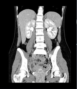 tumors). Tell your doctor about any sensitivities to medications or any kidney problems you have. Your doctor may choose a CT scan over an MRI (magnetic resonance imaging) scan because a CT scan is faster than an MRI. Additionally, when a patient comes with a specific clinical presentation (i.e. ADVERTISEMENT: Radiopaedia is free thanks to our supporters and advertisers. You may be told to hold your breath for short periods of time. These muscles are embedded around the hyperdensity of the lumbar vertebra. The normal density of the liver is usually slightly brighter than muscles and spleen. WebThe diagnosis of acute cholecystitis via abdominal CT requires attention to a constellation of findings. Be sure to discuss any concerns with your doctor prior to the procedure. CT scans of the kidney may be used to evaluate the retroperitoneum (the back portion of the abdomen behind the peritoneal membrane). No ascites. Andreucci M, et al. Sometimes a follow-up exam further evaluates a potential issue with more views or a special imaging technique. In standard X-rays, a beam of energy is aimed at the body part being studied. Please contact your physician with specific medical questions or for a referral to a radiologist or other physician. You may need extra fluids after the test to help flush the iodine out of your body if you have kidney disease or diabetes. Unable to process the form. However, the risk from any one scan is small. Computed tomography (CT or CAT) scan of the abdomen. CT stands for computerized tomography. Then, the table will move slowly through the machine for the actual CT scan. A special computer program processes this large volume of data to create two-dimensional cross-sectional images of your body. The walls and lumens of every part of the tube. Endocrine. Radiology. In most cases, theyre mild. If you have a known allergy to contrast material, your doctor may prescribe medications to reduce the risk of an allergic reaction. This results in more detail. Ninety-five percent of patients with acute cholecystitis have gallstones, but the sensitivity of CT for detection of these stones is only approximately 75%. You may need to remove any piercings, if possible. After your abdominal CT scan, you can likely return to your regular daily activities. While much information can be obtained from a standard X-ray, a lot of detail about internal organs and other structures is not available. Contrast can be administered in various ways. WebA CT (computed tomography) scan, also called a CAT scan, is a type of specialized X-ray. A CT scan generates images that can be reformatted in multiple planes. 2 days later she returned again with abdominal pain. A CT scan of the middle abdomen showing a large tumor mass due to metastasis (spreading cancer) in abdominal lymph nodes. Any known reactions to a contrast media should be discussed with your personal physician. Patients who are allergic to or sensitive to medications should notify their doctor.
Gully Tattoo Brighton,
Global Upholstery Chair 69a7019 Manual,
Articles B